Molecule Tutorials - Herong's Tutorial Examples - v1.26, by Herong Yang
Protein Visualization - Ribbon Diagram
This section provides a quick introduction of protein ribbon diagrams, which uses flat ribbon arrows for beta sheets, and twisted ribbon for alpha helices.
What Is Ribbon Diagram? - A ribbon diagram is a diagram to visualize a protein conformation using ribbons and wires to represent its secondary structures in 3-dimensions.
Here some common conventions used in protein ribbon diagram:
- Flat Ribbon Arrow - Represents a section of protein sequence in a beta sheet structure. The arrow indicates the C-terminal direction.
- Twisted Ribbon - Represents a section of protein sequence in an alpha helix structure.
- Bended Wire - Represents a section of protein sequence in in a loop or turn structure.
The picture below provides a good illustration of a protein ribbon diagram (source: uvm.edu). Note that two small molecules are included in the diagram showing how they are bonded (enclosed) in empty spaces inside the protein.
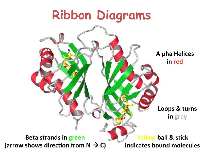
Table of Contents
Molecule Names and Identifications
Peptide, Peptide Bond, Amino Acid Residues
►Protein Visualization - Ribbon Diagram
Composed Proteins or Protein Complexes
wwpdb.org - Worldwide PDB (Protein Data Bank)
Nucleobase, Nucleoside, Nucleotide, DNA and RNA
ChEMBL Database - European Molecular Biology Laboratory
PubChem Database - National Library of Medicine
INSDC (International Nucleotide Sequence Database Collaboration)
HGNC (HUGO Gene Nomenclature Committee)